Angle-Closure Glaucoma Background
Glaucoma is a diverse group of disorders characterized by elevation of the intraocular pressure (IOP) with progressive damage to the optic nerve. Primary glaucoma is not associated with other diseases, while secondary glaucoma is caused by some other ocular abnormality or systemic disease.
An anatomic classification system also is used to describe this group of disorders as closed-angle or open-angle glaucoma. Open-angle glaucoma results in chronic progressive vision loss but generally causes no acute symptoms. In contrast to open-angle glaucoma, acute closed-angle glaucoma (ie, narrow-angle or angle-closure glaucoma) is a true ocular emergency that requires immediate diagnosis and treatment to prevent permanent visual impairment and to relieve pain.
Frequency: In the US: Incidence is approximately 100 cases per 100,000 population.
Race: This condition is more common in African Americans and Asians than whites.
Sex: Females are affected more often than males.
Age: Predominant age range is 55-70 years.
History:
- Patients with an acute attack of closed-angle glaucoma usually present with sudden onset of blurred vision and eye pain.
- Nausea and vomiting also may be present due to vagal stimulation, and some patients may lack severe eye pain that usually is described.
- Diagnosis may be elusive because of systemic symptoms that may mimic cardiovascular or intraabdominal disease. Acute angle-closure glaucoma may be misdiagnosed as a migraine headache, particularly in those with a history of migraine.
- The patient may see halos around lights due to corneal edema.
- Dull ache in or around one eye may be present. Pain is typically deep and boring.
- Symptoms occur when watching TV or movies in a dark room, reading, or when fatigued. Rest often relieves symptoms.
- Acute symptoms: Blurred vision, Severe pain, Lacrimation, Iris bombe, Frontal headache, Nausea and vomiting
Physical:
- Physical examination: , Examination reveals decreased visual acuity. , Conjunctival injection is present, most prominent at the limbus. , The cornea appears hazy from edema. , Ischemia to the iris produces a fixed midposition pupil.
- Subacute: , Normal IOP (10-23 mm Hg) , Shallow anterior chamber , Intermittent peripheral anterior synechiae, which can only be seen by gonioscopy , Enlarged pupil
- Acute: , Elevated IOP (usually 40-80 mm Hg) , Corneal microcystic edema , Lid edema, conjunctival hyperemia, and circumcorneal injection , Fixed, middilated pupil, often oval (may have a relative afferent pupillary defect on swinging light test) , Shallow anterior chamber, often with inflammatory reaction (flare more prominent than cellular reaction)
- Chronic: , Multiple peripheral anterior synechiae , Normal or elevated IOP , Increased cup-to-disc ratio , Normal pupil
Causes: Predisposing ocular anatomy
Lab Studies: No lab studies are needed.
Imaging Studies: No imaging studies are needed.

Contact Us

Manhattan Office
49 West 127th Street
New York, N.Y. 10027
Phone: (212)663-0473
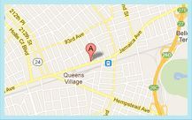
Queens Office
215-43 Jamaica Ave.
Queens Village, N.Y. 11428
Phone: (718) 217-0424