Optic Nerve
For these reasons, examination of the optic nerve and its ability to transmit the visual message is an essential portion of the examination for glaucoma. In most patients, the optic nerve can be readily examined. It can be seen directly inside the eye with an instrument called the ophthalmoscope.
The optic nerve exits through the back of the eye. The nerve is made up of fibers which originate in nerve cells located in the retina, the light-sensitive film coating the inside of the eye. In the normal state, the optic nerve head looks much like a doughnut, with the outer ring consisting of the nerve tissue.
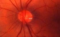
Above is a normal optic nerve head with thick outer ring of nerve tissue. The optic cup is small.
The hole (called the optic cup) is the space which remains after the nerve fibers turn to fan out into the retina. In glaucoma, the nerve fibers are damaged and erode away, leaving a larger cup (or hole of the doughnut).
Especially when the degree of enlargement is different between the two eyes, the physician can diagnose early glaucoma from this appearance alone.
New methods of detecting damage to the optic nerve appearance using computers are being developed, but standard examination with conventional instruments can often detect even early damage. Newer methods that may permit even earlier detection also are being developed to examine and measure the nerve fibers as they spread into the retina.
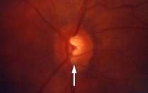
Above is a glaucomatous optic nerve head. The arrow corresponds to the enlarging cup with notching present and loss of the nerve fibers.
Nerve Fiber Laser Analysis
This is new technology that allows us to detect any loss of the nerve fibers from the eye from glaucoma. This often precedes visual field loss. This is a simple test performed in the office.

It is an excellent device for early detection and can monitor early glaucoma even before visual field loss. Above is a picture of the machine:
Below is picture of the normal bimodal nerve fiber pattern in the eye:
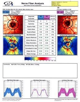

Below is a picture of a patient with glaucoma with a flattened nerve fiber appearance indicating loss of vision:


Contact Us

Manhattan Office
49 West 127th Street
New York, N.Y. 10027
Phone: (212)663-0473
Queens Office
215-43 Jamaica Ave.
Queens Village, N.Y. 11428
Phone: (718) 217-0424