Other Tests
- Tonometric testing reveals increased IOP, the hallmark of glaucoma. Increased IOP can be measured in the ED with a variety of tonometric devices.
- The Schiötz tonometer is the most widely used instrument for measuring intraocular pressure. Schiötz tonometry readings indicating the amount of corneal indentation produced by a standard weight. Scale reading can be converted to an absolute pressure in mm Hg.
- Unfortunately, application of the Schiötz tonometer to the eye often produces an iatrogenic corneal abrasion, particularly if the patient moves the eye during the examination. The appropriate weight must be used to obtain an accurate reading. If the needle of the scale reads at either end of the spectrum (high or low), another size weight should be tried to obtain a reading toward the middle of the scale and the appropriate table for that weight must be used.
- The Goldman applanation tonometer is attached to the slit lamp and measures the force needed to flatten an area of the cornea. Its use requires some training in interpreting the semicircular fluorescein rings seen through the biomicroscope. A boggy or edematous cornea can give a spuriously low reading.
The Tono-Pen is an electronic hand-held applanation tonometer with a tip that is covered by a disposable cover to prevent transmission of ocular infection. This device allows the clinician to use a minimally invasive approach to obtain a rapid reliable reading. One must appropriately calibrate the Tono-Pen before use.
Once diagnosed, immediate treatment is necessary to prevent the rapid ocular damage caused by very high IOP.
Once the pressure is high enough to interfere with arterial or venous flow through the zone of high pressure, the outcome is more dependent on duration of the attack than on the absolute pressure.
Strategies to lower IOP
- Block aqueous production
- Reduce vitreous volume
- Facilitate aqueous outflow
Physical:
- Topical beta-adrenergic antagonists such as timolol and betaxolol decrease aqueous production. Systemic absorption does occur, and these agents should be used carefully in patients with reactive airway disease, heart failure, or cardiac conduction blocks. Topical timolol causes IOP to fall in 30 minutes with peak effects in 1-2 hours. A reasonable regimen is Timoptic 0.5%, one drop every 30 minutes for 2 doses.
- The carbonic anhydrase inhibitor, acetazolamide, also decreases aqueous production and should be given in conjunction with topical beta-antagonists. An initial dose of 500 mg is administered followed by 250 mg every 6 hours. This medication may be given orally, intramuscularly, or intravenously.
- Apraclonidine is a relatively new alpha 2-agonist that acts primarily by decreasing aqueous production. Its effects are additive to topically administered beta-blockers. It has been approved for use in controlling an acute rise in pressure following anterior chamber laser procedures, but has been reported effective in treating acute closed-angle glaucoma. A reasonable regimen is 1 drop every 30 minutes for 2 doses.
Reduce vitreous volume
- In addition to curtailing aqueous production, reduction of vitreous volume with systemic hyperosmotic agents often is needed to break the acute attack. These agents draw water out of the globe by making the blood hyperosmolar.
- Oral glycerol in a dose of 1 mL/kg in a cold 50% solution (mixed with lemon juice to make it more palatable) often is used. Glycerol is converted to glucose in the liver; persons with diabetes may need additional insulin if they become hyperglycemic after receiving glycerol.
- Oral isosorbide is a metabolically inert alcohol that also can be used as an osmotic agent for patients with acute angle-closure glaucoma. Usual dose is 100 g taken PO (220 cc of a 45% solution). This inert alcohol should not be confused with isosorbide dinitrate, a nitrate-based cardiac medication used for angina and for congestive heart failure.
- Intravenous mannitol in a dose of 1.0-1.5 mg/kg also is effective and is well tolerated in patients with nausea and vomiting.
- These hyperosmotic agents should be used with caution in any patient with a history of congestive heart failure.
Facilitate aqueous outflow
- Facilitation of aqueous outflow is the third step in reducing IOP. This can be accomplished by instilling a drop of pilocarpine 2-4% every 15 minutes for the first 1-2 hours. This miotic agent pulls the iris from the iridocorneal angle and may help to relieve the obstruction of the trabecular meshwork by the peripheral iris. More frequent administration or higher doses may precipitate a systemic cholinergic crisis.
- Because the effect of pilocarpine varies depending on the color of the iris, use of the 4% solution on brown and 2% on blue irises has been advocated. The IOP usually needs to be brought below 40 mm Hg, the perfusion pressure of the iris, before pharmacologic miosis can be achieved.
- Because the goal is to lower IOP as quickly as possible treatment with one of the osmotic agents, as well as acetazolamide, pilocarpine, topical beta-blocker, and alpha 2-agonist should be performed concurrently.
- Once the acute attack has been broken, the definitive therapy for narrow-angle glaucoma is surgical. A peripheral iridotomy, surgical or by laser therapy, is performed. The hole provides an alternative pathway for aqueous humor to reach the anterior chamber and relieves the relative pupillary block. Because the contralateral eye has the same anatomy, predisposing it to an acute attack of closed-angle glaucoma, a prophylactic iridotomy in that eye also is performed.

Contact Us

Manhattan Office
49 West 127th Street
New York, N.Y. 10027
Phone: (212)663-0473
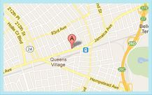
Queens Office
215-43 Jamaica Ave.
Queens Village, N.Y. 11428
Phone: (718) 217-0424